
The most commonly affected body system by unhealthy lifestyle choices like smoking, lack of exercise, and fatty food intake is the cardiovascular system. This comprises the heart and your blood vessels. Over time, your lifestyle leads to the deposition of plaque along the walls of your blood vessels. This narrows the vessels and leads to hypertension. The plaque can also detach from the vessels’ walls and block your pulmonary artery, effectively cutting off blood supply to the heart.
The most common test done at a diagnostic clinic based in London to assess the condition of your veins and arteries is an angiography. This uses real-time image guidance to generate images of your blood vessels and takes an hour or two. Catheters are placed into your blood vessels, and then a radio-opaque contrast agent is injected into them. As the dye passes through a vessel, images will be conveyed to a screen from where a specialist will make a diagnosis of its condition. The following are the angiography types that a specialist might order:
Coronary Angiographies
This is done on your heart arteries to pick sections of narrowing that might cause heart attacks in the future. The narrowed arterial parts are also often the common causes of sharp chest pain. The tip of a catheter is generally placed into a vessel in your groin or arm from where a dye is injected. Coronary angiography can also be used for interventional procedures when a valve or drug is introduced into the blood vessels through the catheter.
CT Angiography
This uses x-rays for the generation of clear, detailed images of your body tissues. In coronary CT (computed tomography) angiography, for instance, a doctor will pick issues with your heart valves and muscles, aorta, and the pericardium (the sac around your heart). The test also detects clots in your lungs. In CT angiography, the generated images resemble thin cross-sections of the area of interest from different angles.
Optical Coherence Imaging
This is a high-resolution diagnostic imaging technique. It, unlike other angiographies, will use somewhat infrared light to get microscopic images of your coronary arteries. Optical coherence imaging is used for the assessment of narrowed arteries and the effect of stent placement during a percutaneous coronary therapeutic intervention.
Cerebral Angiography
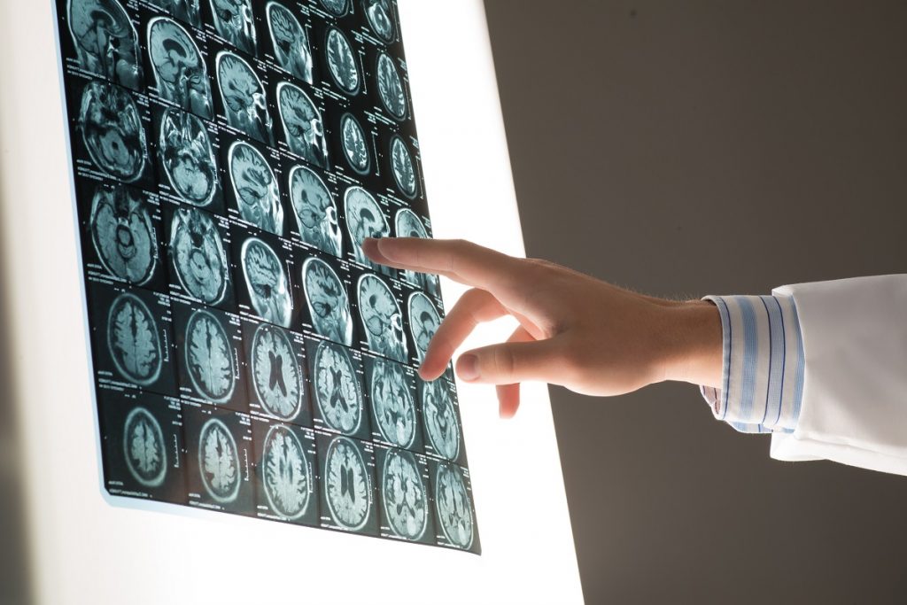
This will assess the blood vessels supplying blood to your brain and draining the blood from it. Any blockages in these blood vessels might cause a stroke that affects your body in varying degrees. Using cerebral angiography, your doctor can determine your risk of getting a stroke and intervene early and appropriately.
Renal Angiography
This evaluates the condition of blood vessels supplying your kidneys. When blocked, you risk the insufficient cleaning of the wastes in your body. The catheter used for the angiography can also be used to deliver drugs to the kidneys and insert stents to open them if blocked.
Compared to most procedures, the above angiographies are relatively fast. Though painless, a doctor can use a local anesthetic on the tip of a catheter. This minimizes the discomfort you experience during the catheter’s insertion.